Question
Question: The final image formed by a compound microscope is: A. Real and erect B. Virtual and erect C. ...
The final image formed by a compound microscope is:
A. Real and erect
B. Virtual and erect
C. Real and inverted
D. Virtual and inverted
Solution
We know that a compound microscope consists of two lenses that work together to bring about appropriate magnification. Recall that the role of the objective lens is to ensure that the image is placed in just the right place for the eyepiece to magnify the image while retaining good clarity. Usually, the object is placed just beyond the objective lens. In such a case, determine the kind of image formed, keeping in mind that this serves as the object for the eyepiece, following which you can arrive at the nature of the image consequently formed.
Complete step-by-step answer:
We know that a compound microscope is an optical device using which we can obtain large values of magnification. It is made up of two convex lenses placed along the optic axis: Objective lens and Eyepiece. Usually, the focal length of the objective lens is less than the focal length of the eyepiece since the magnifying power of a compound microscope is inversely proportional to the focal length of the objective lens. The eyepiece is essentially used to magnify the image to a scale where it is observable by the human eye, whereas the objective lens is responsible for placing the image in the right place for the eyepiece to magnify the image with a good clarity and resolution.
Converging lenses in general, can produce both real and virtual images. The image is said to be real if it is formed by the actual convergence of rays, whereas it is said to virtual if it is formed by the convergence of the extensions of diverging rays. An easy way to distinguish the two is to see if the image formed is on the same side of the lens as the object or not. If it’s on the same side then the image is likely to be a virtual one, else it is a real image.
Converging lens can produce both inverted and upright images. An image is said to be inverted if it has a vertical orientation opposite to that of the object, this means that the image is upside-down compared to the object. But if it is vertically oriented in the same way as that of the object, then the image is said to be upright.
The image formed by the lens thus varies based on the position of the object from the lens.
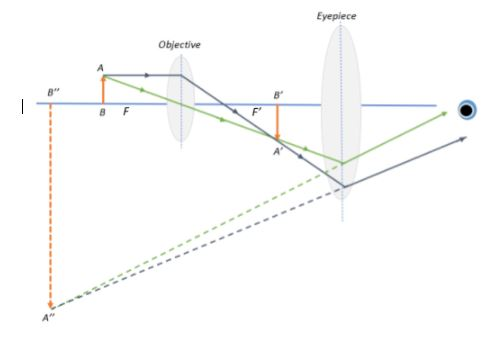
Now, from the figure, we can see that the object is placed just beyond the focal point F. The image formed by the objective lens is real and inverted, since the image is on the opposite side of the lens to the object and is oriented opposite to the object in the vertical direction.
The image formed by the objective lens now serves as the object for the eyepiece and is located between its focus F’ and its centre. The image formed is on the same side as that of the object and is hence, virtual, and since the image is oriented opposite to the object in the vertical direction, it is still inverted.
So, the correct answer is “Option D”.
Note: The above problem does not discuss the magnification of the image. Also, it is a common mistake to interchangeably use the terms enlarged and magnified. Note that enlarged means that the image is bigger relative to the object, whereas magnified can imply either an enlarged image or a diminished (minified) image. This is usually given by:
Magnification M=Object heightImage height
If ∣M∣<1 then the image is diminished, and if ∣M∣>1 then the image is enlarged. If ∣M∣=1 it means there is no magnification and the image is the same size as that of the object.
The ‘+’ sign indicates that the image is upright, whereas the ‘-’ sign indicates that the image is inverted.
Thus, from the question, we see that the final image height is much greater than the object height, i.e., ∣M∣>>1. And since the final image is inverted, the magnification of the compound microscope will be −∣M∣