Question
Question: In a compound microscope the lens used are A. Both convex of same focal length B. both convex of...
In a compound microscope the lens used are
A. Both convex of same focal length
B. both convex of different focal length
C. both concave of same focal length
D. one convex and one concave of different focal length.
Solution
A compound microscope is an optical device which produces magnified images of tiny objects. A good quality compound microscope can produce magnification in order of 1000. In compound two lenses were used so that the combined magnification of the compound microscope is increased and a much magnified image of the tiny object is seen in the eye piece. The length of two lenses used is different. The objective (the lens near the object) is of small size whereas the eyepiece (the lens near the eye) is of different size.
Formulas used:
Magnification of compound microscope is given by.
M=θ0θ1
Where θ1 and θ0are the angle subtended by image and object at the eyepiece.
Magnification of simple microscope of focal length f and distance of image from the lens D is
M=1+fD
Complete answer:
The convex lens ‘O’ of short focal length and small aperture, which faces the object to be viewed, is known as the objective. The convex lens ‘E’ of large focal length and comparatively large aperture, near the eye, is known as eyepiece. The two lenses are placed in two metal tubes so as to have a common principal axis. The eyepiece is fitted in a draw-tube and can be slided within the main tube by means of rack and pinion arrangement to focus the microscope upon the object.
Let AB be an extended object situated on the principal axis at a distance slightly greater than the focal length of the objective. As refraction takes place through the objective ‘0’, a real, inverted and magnified image A1B1 is formed. The lens E is so adjusted that A1B1 falls within its focal length and so the final virtual image A2B2of A1B1 is obtained at the least distance of distinct vision 'D' from the eye. The final image A2B2 is thus highly magnified but is inverted with respect to the object AB.
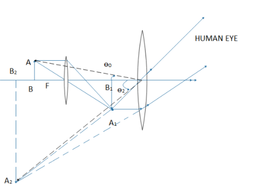
When the final image is obtained at the least distance of distinct vision from the eye.
The magnifying power of a compound microscope is defined as the angle subtended by the final image at the eye to the angle subtended by the object at the eye when both the object and the image are situated at the distance of distinct vision from the eye.
If the object is situated at B2, it would have occupied the length such that B2C=AB.
Let the angles subtended by the object B2Cand final image A2B2at the eye be θ0 and θ1 respectively
Then the magnification of the compound microscope will be
M=θ0θ1=tanθ0tanθ1(∵ because θ1andθ0 are very small)
In triangle O2B2C, tanθ0=O2B2B2C=O2B2AB
In triangle O2B2A2, tanθ1=O2B2A2B2
Now magnification
M=tanθ0tanθ1=O2B2ABO2B2A2B2=ABA2B2=A1B1A2B2×ABA1B1
But the magnification of objective is M1=ABA1B1 and that of eyepiece is M2=A1B1A2B2
So, M=M1×M2
For objective lens
Magnification=distance of objectdistance of image=uv⇒M1=uv
Also the eyepiece acts like a simple microscope so its magnification is given by M2=1+feD
Where fe= focal length of eyepiece
Now the magnification of compound microscope is given by
M=uv(1+feD)
If the object AB is situated very near the principal focus of the objective, the image A1B1will be far removed from the lens ‘O’. In that case
u=f=focal length of the objective
v=L=length of the microscope tube.
So magnification of compound microscopes now becomes.
M=f0L(1+feD)
So in order to have large magnification the objective lens and eyepiece lens should have small focal length.
In a compound microscope the length of objective is less than the length of eyepiece. So the focal length of the lens should be smaller than the eyepiece.
So the correct option is. B.
Note:
The resolving power of a compound microscope is the inverse of the distance between the two points that can be resolved. For a high resolving power the refractive index must be more. For a good microscope the magnification must be large. In a compound microscope the combination of lenses are used to get a good quality image.