Question
Question: How are the DNA fragments separated by gel electrophoresis visualized and separated for use in const...
How are the DNA fragments separated by gel electrophoresis visualized and separated for use in constructing recombinant DNA?
Solution
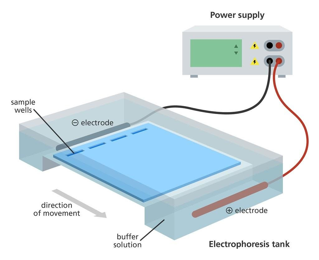
Gel electrophoresis is a technique used to separate the charged particles or molecules on the basis of their molecular size under the influence of electric field. Mixtures of DNA, RNA and proteins can be separated by this technique. This technique was developed by Tiselius in 1937.
Complete answer:
Gel electrophoresis is a technique for separating DNA fragments based on their molecular size.
Firstly, the DNA sample is cut into fragments by restriction endonucleases.
The negatively charged DNA fragments can be separated by letting them move towards the positive charge (anode) under an electric field by the help of a matrix.
Matrix that is used in this technique is agarose, which is a natural linear polymer of D-galactose and 3,6-anhydro-L-galactose which is extracted from sea weeds.
Then, the DNA fragments are separated out according to their molecular size with the help of sieving property of agarose gel.
Small size fragments move fast as compared to large size fragments.
The separated DNA fragments are visualized only after staining the DNA with the help of ethidium bromide followed by the exposure to UV radiation.
The bright orange colour bands are shown.
Then the elution is done, that is the separated bands of DNA are cut out from the agarose gel and extracted from the gel piece.
The purified DNA fragments are used to form recombinant DNA which can be joined with cloning vectors.
Note:
Cloning vectors are the DNA molecules that can carry a foreign DNA segment into the host cell. A technique which is used to change the phenotype of an (host) organism when a genetically altered vector is introduced and integrated into the genome of the organism. The recombinant gene is the gene which is introduced and this technique is termed as Recombinant DNA technology.