Question
Question: Describe giving diagrams of the structure of the eye and the process of vision....
Describe giving diagrams of the structure of the eye and the process of vision.
Solution
Eyes are responsible for vision. It converts light into electrical signals. Eyes are made up of components named as the cornea sclera, pupil, iris, lens retina optic nerve, photoreceptors rods and cones, aqueous and vitreous humour
Complete answer:
The eye is the organ of sight or vision and consists of the following components:
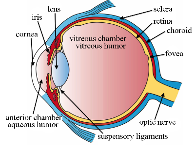
Cornea: Front window of the eye that performs the function of transmitting and focusing light inside the eye.
Sclera: Outer white covering of the eye.
Pupil: Black dot present in the middle of the eye.
Iris: Coloured area of the eye surrounding the pupil. It controls the amount of light entering the eye. In a darker environment iris constricts the pupil to allow more light and in brighter regions, iris dilates the pupil decreasing the amount of light entering the eye.
Lens: Behind iris lens is present. It focuses light on the retina by changing shapes with the help of ciliary muscles.
Retina: It contains photoreceptors and blood vessels. Retina has a region compactly packed with photoreceptors known as macula and this is the most sensitive part of the eye.
Optic nerve: Nerve fibres from photoreceptors group together to form the optic nerve. Photoreceptors: These are responsible for conversion of an image into electrical signals which are transferred to the brain by the optic nerve. Two types of photoreceptors are
Cones: For coloured sharp central vision and Rods: For night and peripheral vision
Aqueous and vitreous humor: Anterior portion of the eye from the cornea to front of the lens is filled by aqueous humour and the posterior segment from to the retina is filled with vitreous humour.
Mechanism of vision: Light after bending from the object falls on the cornea and passes through the lens. These light rays are focused by the lens onto the retina. The retina contains photoreceptors rods and cones which contain photopigments such as retinal and opsin. Light causes retinal and opsin to disassociate leading to structural changes in opsin which in turn changes the permeability of retinal cells which generate an action potential. This action potential is transferred by the optic nerve to the brain where the image is recognized.
Note: The pupil of the eye adjusts its size according to the intensity of light. It dilates in the presence of low light and contracts or shortens in the bright light. This helps in proper vision.